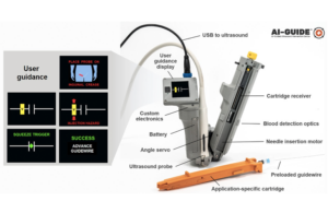
Researchers at the Massachusetts Institute of Technology’s Lincoln Laboratory have developed an artificial intelligence-guided ultrasound device to help physicians quickly deploy a catheter at a point of injury.
Following traumatic accidents, medical professionals often have to apply life-saving treatment to patients with severe internal bleeding. Delivering catheters into a central blood vessel to administer fluid or medication can be complex, the researchers said, and first responders like EMTs are not really trained to perform those types of procedures. Oftentimes, treatment can only be administered at the hospital.
The team of researchers, led by Laura Brattain and Brian Telfer from the Human Health and Performance Systems group, along with physicians from the Center for Ultrasound Research and Translation at Massachusetts General Hospital, developed a solution to the problem. Its AI-guided ultrasound interventions device (AI-GUIDE) is a handheld platform technology that could help physicians with training to quickly install a catheter into a common femoral vessel to enable the rapid treatment at the point of injury, according to the researchers.
“Simplistically, it’s like a highly intelligent stud-finder married to a precision nail gun,” Matt Johnson, a research team member from the Laboratory’s Human Health and Performance Systems Group, said in a news release.
AI-GUIDE is designed with custom-built algorithms and integrated robotics that are able to be paired with most commercial portable ultrasound devices. To operate the handheld surgical robot, a user places it on a patient’s body near where the thigh meets the abdomen and a targeting display guides the user to the correct location and instructs them to push a button that precisely inserts the needle into the vessel, the researchers said.
The device is able to verify that the needle has penetrated the blood vessel and tells the user to advance a guidewire to guide a catheter into the vessel. The physician can then manually advance the catheter. Once the catheter is securely in the blood vessel, the device withdraws the needle and the user can remove the device. The catheter then delivers fluid, medicine and other interventions after it is safely inside the vessel.
“Using transfer learning, we trained the algorithms on a large dataset of ultrasound scans acquired by our clinical collaborators at MGH,” said Lars Gjesteby, a member of the Laboratory’s research team. “The images contain key landmarks of the vascular anatomy, including the common femoral artery and vein.”
The algorithms are able to interpret the visual data coming from the ultrasound that is paired with AI-GUIDE and can indicate the correct blood vessel location to the user on the display.
“The beauty of the on-device display is that the user never needs to interpret, or even see, the ultrasound imagery,” said Mohit Joshi, the team member who designed the display. “They are simply directed to move the device until a rectangle, representing the target vessel, is in the center of the screen.”
The researchers tested AI-GUIDE on an artificial model of human tissue and blood vessels, as well as on a series of live, sedated pigs. The researchers said that all users of the device on artificial human tissue were successful in placing a needle within two minutes of verbal training.
“AI-GUIDE has the potential to be faster, more precise, safer, and require less training than current manual image-guided needle placement procedures,” Massachusetts General Hospital radiologist Dr. Theodore Pierce said.
The team plans to continue testing the device and will work on fully automating the operation of the device. They also want to automate the guidewire and catheter insertion steps to further reduce the risk of user error or infection.
“Retraction of the needle after catheter placement reduces the chance of an inadvertent needle injury, a serious complication in practice which can result in the transmission of diseases such as HIV and hepatitis,” said Pierce. “We hope that a reduction in manual manipulation of procedural components, resulting from complete needle, guidewire, and catheter integration, will reduce the risk of central line infection.”

