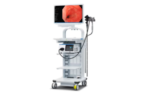
The FDA clearance covers Olympus’ GIF-1100 gastrointestinal videoscope indicated for use in the upper digestive tract, and the CF-HQ1100DL/I colonovideoscope indicated for use in the lower digestive tract.
Olympus’s GI endoscopy systems are used by physicians to help diagnose, treat and observe diseases and disorders of the upper and lower GI tract, such as acid reflux, ulcers, Crohn’s disease, Celiac disease and colorectal cancer. According to the company, one of the most common uses of an endoscope is for screening colonoscopy, during which a physician examines the lining of the colon and removes cancerous growths. Olympus’ new imaging technologies can help physicians visualize abnormalities.
“We are thrilled that we will soon be able to bring this new endoscopy system to physicians and their patients in the U.S.,” Richard Reynolds, president of the medical systems group for Olympus America, said in a news release. “As a leading medical technology company, Olympus strives to offer physicians the most advanced technologies for minimally invasive procedures such as GI endoscopy.”
More about the endoscopy system
The Evis X1 endoscopy system has three enhancements that the company designed to assist physicians in visualizing GI bleeds and anatomical structures. The technology is enabled by the replacement of the Xenon bulbs in the Evis Exera III system with five LEDs that can produce other light combinations in addition to white light.
Other features include:
- Red dichromatic imaging (RDI) technology for optical-digital observation using red dichromatic narrow band light and green illumination light.
- Texture and color enhancement imaging (TXI) technology to emphasize tonal changes, patterns and image outlines and correct the brightness of dark areas.
- Brightness adjustment imaging with the maintenance of contrast (BAI-MAC) maintains the brightness of the bright part of the endoscopic image and corrects the brightness of the dark part of the endoscopic image.
Olympus’ optical-digital technology NBI (Narrow Band Imaging) technology is also used in the Evis X1 system. It enhances the observation of mucosal tissue and works by filtering white lite into specific light wavelengths that are absorbed by hemoglobin and penetrate only the surface of human tissue. Because of that, capillaries on the mucosal surface appear brown and veins in the submucosa appear cyan on the monitor.

