The prospective, randomized (2:1 treatment to control) dual-center study plans to enroll up to 60 subjects. Investigators intend to use optical coherence tomography (OCT) imaging to demonstrate the mechanism of action (MOA) of Serranator. The study compares the system’s MOA to conventional angioplasty across a range of lesion morphologies in below-the-knee arteries.
Co-principal investigators Dr. Sahil Parikh and Dr. Brian DeRubertis lead the study. The company selected sites at Columbia University Medical Center and Weill Cornell Medicine. It says this marks the first study of its kind: randomized, utilizing OCT imaging to compare acute outcomes between serration angioplasty and POBA.
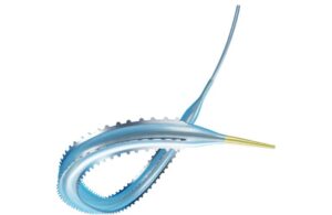
Serranator, an FDA-cleared, novel balloon, uses stainless steel micro-serration technology. Cagent designed it to create linear, interrupted scoring along the endoluminal surface. It features 1,000 times more point force compared to POBA, delivering serration during slow, low balloon inflation. It aids arterial expansion, effectively achieving luminal gain in all lesion morphologies with minimal recoil.
Parikh says the study can help emphasize guidance on what interventions may be most appropriate for Cagent’s Serranator technology.
“OCT provides a novel visualization of vascular disease and therapeutic results. With 10x the resolution of IVUS, we believe this study will allow us to understand how Serration Angioplasty and Plain Balloon Angioplasty interact with the intima, internal elastic lamina, and media. We’re eager to assess this visually and quantitatively in this first-of-its-kind study,” said DeRubertis.